
Fig. 4.
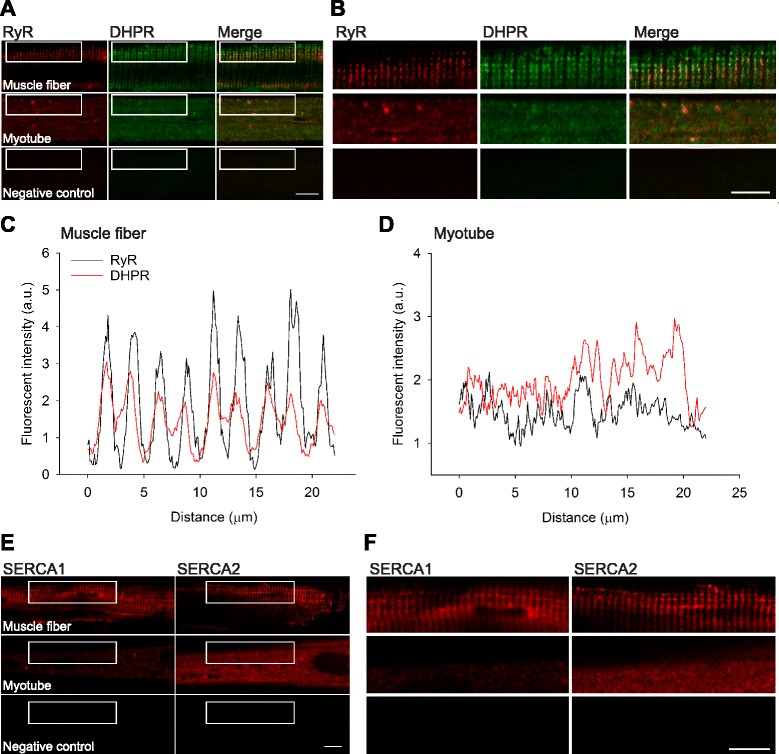
The localization of Ca2+-handling proteins differs markedly between human muscle fibers and myotubes. a Representative immunofluorescent staining for RyR and DHPR in human muscle fibers (top row) and myotubes (middle row). b Magnification of the area indicated in a. c, d Fluorescent intensity (a.u.) for RyR (black) and DHPR (red) in a human muscle fiber (left) and myotube (right). e Representative immunofluorescent staining for SERCA1 and SERCA2 in human muscle fibers (top row) and myotubes (middle row). f Magnification of the area indicated in e. Scale bars indicate 10 μm. Primary antibody was omitted for the respective negative controls (bottom row in a, b and e, f)