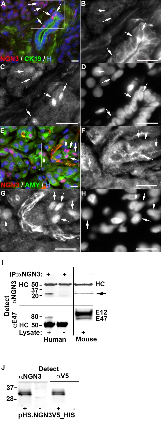
Fig 1. Expression of neurogenin 3 (NGN3) in the adult human exocrine pancreas.

A-H, Immunohistochemical staining of histologically normal tissue from living subjects undergoing medically indicated pancreas biopsy using anti-NGN3 antibody F25A1B3. A-D, Expression of NGN3 and cytokeratin 19 (CK19) in by duct cells. B-D, Higher magnification of tissue shown in A. B, CK19 expression, C, NGN3 expression, D, Hoechst 33342 stained nuclei (H). E-H, Expression of NGN3 and amylase (AMY) by acinar cells. F-H, Higher magnification of tissue shown in E. F, Amylase expression, G, NGN3 expression, H, Hoechst 33342 stained nuclei. NGN3+ cells indicated by white arrowheads. Scale bars are 20 μm. I, Immunoprecipitation (IP) of NGN3 and E12/47 from human exocrine tissue after 4 days of culture. Presence of IP antibody F25A1B3 shown on top. F25A1B3 and anti-E12/47 detection antibodies shown on left. Presence of human or E14.5 mouse pancreatic epithelia lysate shown on bottom. Detection with anti-NGN3 (top panel) reveals bands in human and mouse lysates corresponding to the predicted molecular mass of NGN3 (~23KDa, arrow) and capture antibody heavy chain (HC). Detection with anti-E12/47 (bottom panel) identifies coimmunopreciptated proteins corresponding in size to E12 and E47. Molecular weight markers shown at left of blots in kDa. J, HEK293T cell lysate expressing human NGN3 tagged with V5 and 6xHIS epitopes (pHS.NGN3V5_HIS) (+) or negative control vector (-) detected with NGN3 antibody F25A1B3 and anti-V5 as indicated. Molecular weight markers shown at left of blots in KDa.
