
Fig. 5.
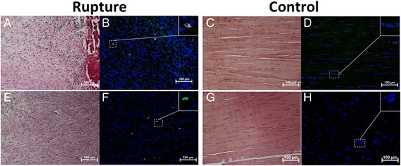
Dynamin 2 and Oct 3/4 in I region in early tendon healing. Sections showing few positive Oct 3/4 (yellow) (b) and Dyn 2 (green) (f) cells inside the I region of Achilles rat tendon at one week post-rupture. No Oct 3/4- (d) or Dyn 2- (h) -positive cells were found inside the I region of control tendons (contralateral intact tendon). Histological staining of sections of ruptured (a,e) and control (c,g) sections was performed on adjacent consecutive sections. Cell nuclei stained with DAPI (blue)