
Fig. 9.
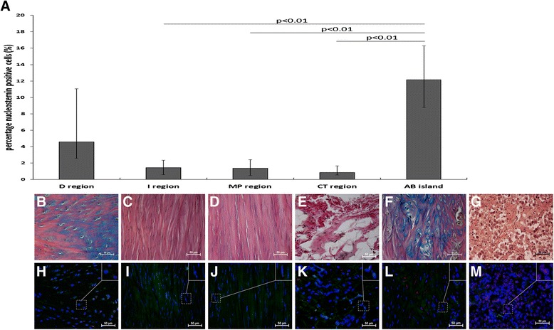
Nucleostemin percentage and presence at different locations in healing tendon at week 17. The percentage of nucleostemin-positive cells was calculated in five different regions from ruptured (right leg; n = 6) tendons at week 17. The diagram (a) shows a significantly higher percentage of nucleostemin-positive cells in regions with high Alcian blue content (AB islands) compared with I, MP and CT regions. Values are expressed as the median with IQR as error bars. Images showing histological staining (b-f) and nucleostemin (red) staining (h-l) in the different regions of the tendons. Positive control tissue for nucleostemin was human seminoma tissue (g,m)