
Fig. 6.
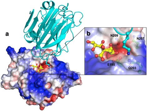
Potential functional cavity of SpCBM. a The surface electrostatic view from the top of the binding site of SpCBM and a cavity adjacent to the active site, which is occupied by a lysine from a symmetry related molecule (shown in cyan). b Close-up view of the interaction between the lysine and the protein. The solvent-accessible surface of SpCBM is coloured based on the electrostatic potential from −7 (red) to +7 (blue) kT/e, calculated using the APBS tool in PyMOL [34, 35]