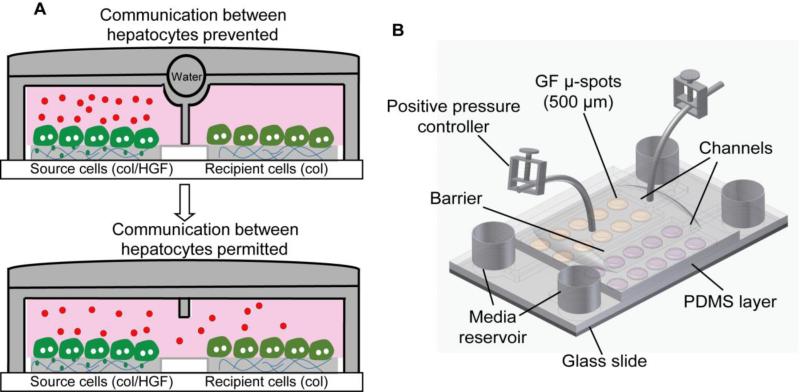
Fig. 1.
Reconfigurable microfluidic device. (A) Schematic showing the communication between two groups of hepatocytes through endogenous signals. Microfluidic chambers in its close state allow accumulation of secreted signals produced by differentiated hepatocytes (source cells) on col/HGF spots in small volume. The accumulated signals can diffuse into next chamber with deactivation of barrier and may control the cellular behavior of neighboring cells (recipient cells). (B) Schematic illustration of a two-layer reconfigurable microfluidic device. The bottom PDMS layer binds to glass and defines the dimensions of the microfluidic channels. The top layer binds to the bottom layer and contains a water chamber that is used to switch between open and closed states of the device. Col, collagen type I.

