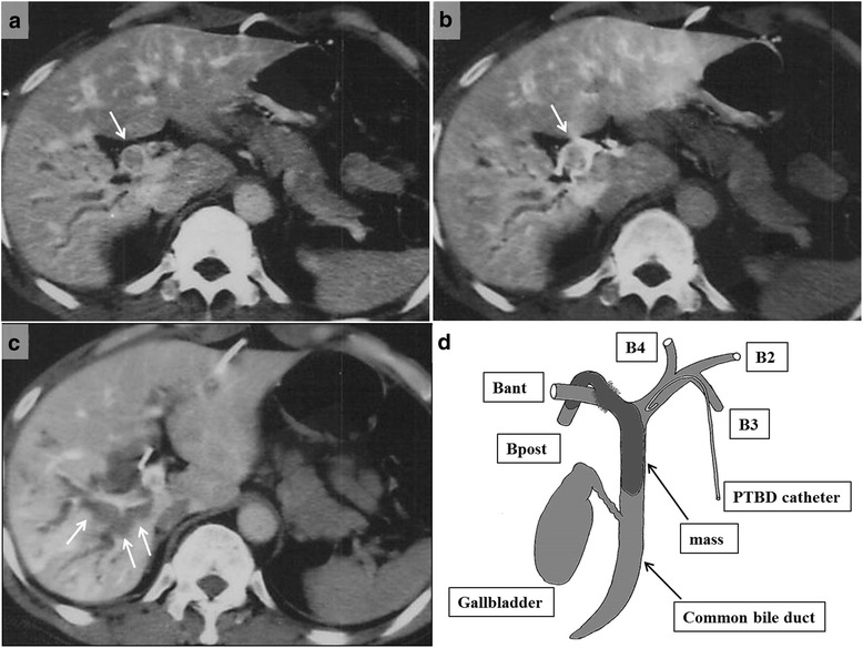
Fig. 3.

Follow-up contrast-enhanced computed tomography 9 years after sigmoidectomy. CT scans revealed a, b intrahepatic bile duct dilatation (single arrow) in the posterior segment of the right lobe to the common hepatic duct and c a low-density mass (arrows) in the dilated bile duct. A schematic diagram (d) shows the tumor location. Bant anterior right hepatic duct, Bpost posterior right hepatic duct, B2 segment 2 bile duct, B3 segment 3 bile duct, B4 segment 4 bile duct
