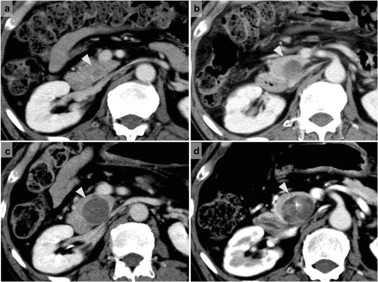
Fig. 8.

Contrast-enhanced computed tomography revealed the enlargement of the tumor in the intrapancreatic bile duct. CT scans obtained in a October 2005 (2 years and 10 months after hepatectomy) depicted a very low-density mass in the intrapancreatic bile duct. CT scans obtained in b December 2007 (5 years after hepatectomy), c January 2011 (8 years and 1 month after hepatectomy), and d February 2014 (11 years and 2 months after hepatectomy) show gradual dilatation of the intrapancreatic bile duct; the mass had gradually enlarged
