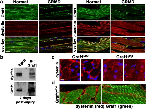
Fig. 8.

GRAF1 associates with dysferlin in injured/dystrophic muscles and promotes its recruitment to the PM. a Immunohistochemical analysis of GRAF1 expression (green) in normal and dystrophic (golden retriever muscular dystrophy; GRMD) muscle. Co-staining with dysferlin (red, left) and α-actinin (red, right) demonstrates localization of GRAF1 in striated pattern aligned with Z-disks in normal muscle compared to localization to the plasmalemma in GRMD muscle (note intramuscular inflammatory infiltrates indicative of muscle injury as visualized by DAPI staining). b Anti-GRAF1 rabbit polyclonal antibody [52] immunoprecipitation (IP) from adult quadriceps muscle 7 days following CTX-induced injury. Blots were probed with an anti-dysferlin antibody or hamster anti-GRAF1 antibody. Input contains 2 % of cellular lysate used for IP. c Immunohistochemical analysis of dysferlin (red) in Graf1Wt/Wt and Graf1Gt/Gt adult mouse hearts indicates dysferlin mis-localization in the absence of GRAF1. Nuclei are counterstained with DAPI (blue). d Immunohistochemical analysis of dysferlin (red) and GRAF1 (green) reveals plasmalemmal and sarcomeric localization respectively in Wt hearts. Scale bars = 20 μm
