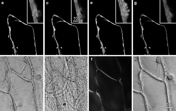
Fig. 1.

The importance of optimised illumination for transmitted light imaging. Onion epidermal cells transiently expressing a cytosolic targeted YFP were imaged by confocal fluorescence microscopy (a, c, e, g) and concurrently with various forms of transmitted light (b, d, f, h). a, b Optimised fluorescence and transmitted light imaging, with the transmitted light path set for Köhler illumination. c, d Non-optimal transmitted light imaging, with the condenser lens defocused and condenser diaphragm stopped down, resulted in a contrasted image with large depth-of-field. e, f Polarised light imaging revealed the location of the cell walls because of cellulose birefringence. g, h DIC imaging improved transmitted light detail, but reduced the brightness and quality of the fluorescence image as shown in the inset images. Scale bars 50 µm for main images and 10 µm for insets
