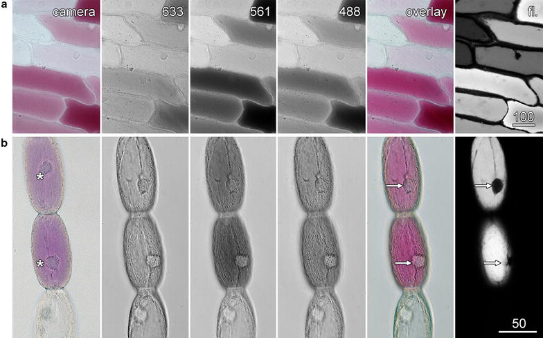
Fig. 2.

Colour images with a monochrome transmitted light detector. Transmitted light images were recorded concurrently with the 633 nm red, 561 nm green and 488 nm blue lasers, with these images pseudocoloured and combined into an overlay image (overlay) which matched the colour camera image (camera). Anthocyanin fluorescence images (fl.) were also collected. Arrows indicate nuclei in the colour transmitted light and fluorescence images, while the asterisks indicate the nuclei in the colour camera image that had moved between confocal and bright-field imaging. a Red onion epidermis. b Tradescantia stamen hair cells. Scale bars 100 µm (a) and 50 µm (b)
