
Fig. 2.
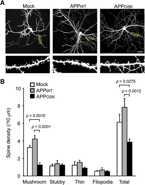
Spine alteration in APPOSK-expressing neurons. a Mouse primary neurons were doubly transfected with APP and GFP. Scale bar, 30 μm. Lower panels, enlarged views of the dendrites surrounded with a square. b Individual spines on dendrites of GFP-positive neurons were classified into the four types: mushroom, stubby, thin, and filopodia-like protrusions. Compared with mock-transfectants, APPWT-expressing cells showed a small increase the number of total and mushroom-type spines with no significant changes in other types. In contrast, APPOSK-expressing cells exhibited a significant reduction in the number of total and mushroom-type spines, but no significant changes in other types