
Fig. 5.
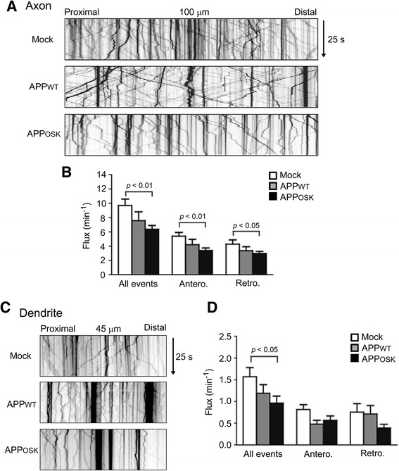
Axonal and dendritic transport of BDNF is impaired in APPOSK-expressing neurons. Wild-type mouse primary neurons were doubly transfected with APP-EGFP and BDNF-mRFP. Transport of BDNF in living neurons was recorded for 25 s, and those in 100-μm segments of the axon (a) and 45-μm segments of the dendrite (c) were analyzed. Axons and dendrites were initially identified based on morphology and confirmed retrospectively by immunostaining MAP2. b Compared with mock-transfectants, both APPWT- and APPOSK-expressing cells showed a decrease of bidirectional transport of BDNF in axons, but the differences were significant only in APPOSK-expressing cells. d Similarly, a decrease of bidirectional transport of BDNF in dendrites was observed in both APPWT- and APPOSK-expressing cells, but significant reduction was observed only for total event in APPOSK-expressing cells