
Fig. 7.
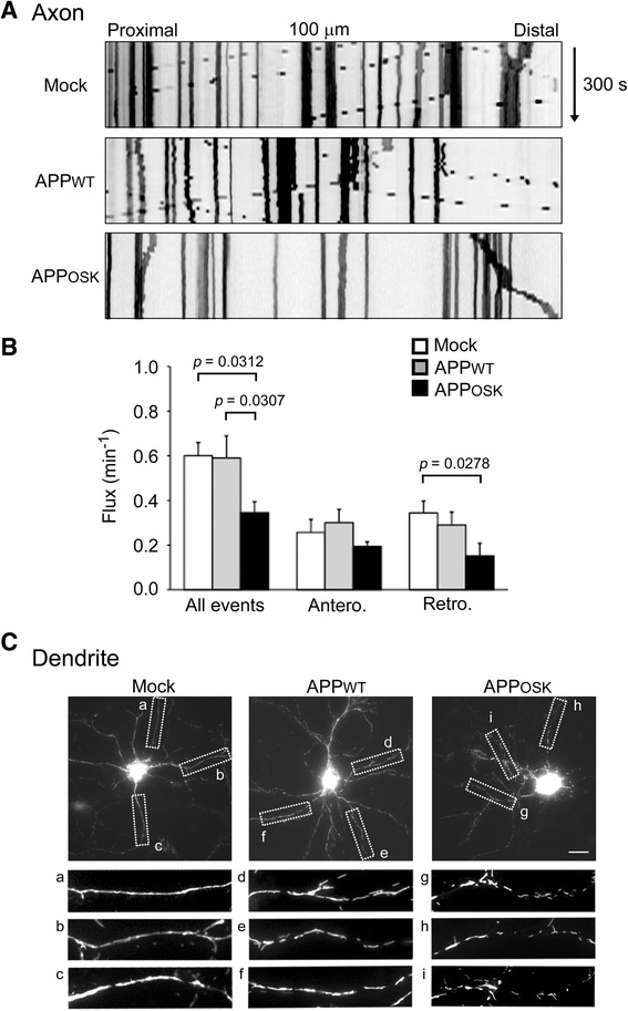
Impaired axonal transport and aberrant dendritic distribution of mitochondria in APPOSK-expressing neurons. Rat primary neurons were triply transfected with APP, Mt-eYFP, and BFP. a Transport of mitochondria in living neurons was recorded for 300 s, and those in 100-μm segments of the axon were analyzed. b Compared with mock-transfectants, bidirectional transport of mitochondria in axons was reduced in APPOSK-, but not APPWT-, expressing cells. c In dendrites, mitochondria were evenly distributed along the dendritic shafts in mock-transfectants (a-c) and APPWT-expressing cells (d-f). In contrast, mitochondria in dendrites of APPOSK-expressing cells (g-i) were of shorter length and unevenly distributed. Scale bar, 20 μm