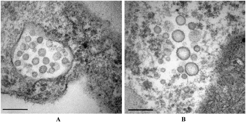
Fig 2. Electron micrograph of a MVBaV-infected mulberry plant leaves showing typical tospovirus-like particle morphology.
Typical spherical, enveloped virions are shown accumulating in the endoplasmic reticulum (A) or dispersing in the cytoplasm as single particles in leaf cells (B). The scale bar represents 200 nm.

