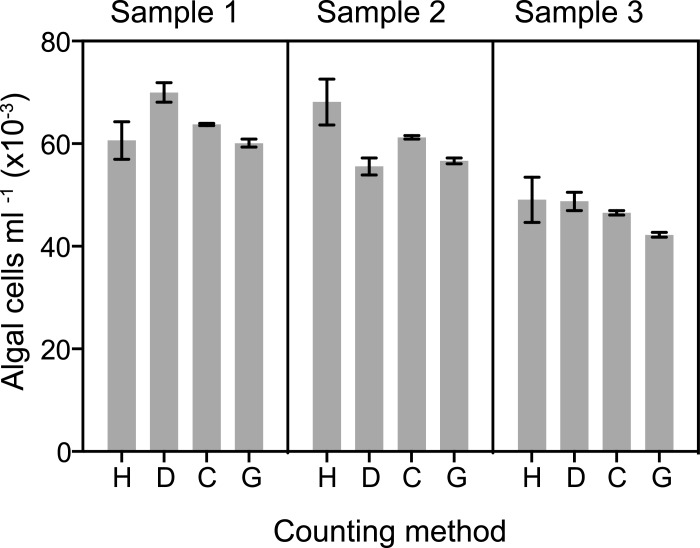
Fig 2. Differences in precision of different methods for counting algal cells.
Symbiotic anemones of strain CC7 were washed in ASW and suspended in a small volume of solution containing one part ASW, 7 parts dH2O, and ~0.08% SDS. The animals were then homogenized in a manual tissue homogenizer (see Materials and Methods), and Samples 1, 2, and 3 were prepared by further dilution of the homogenate with the same solution. The concentration of algal cells in each sample was then determined using a hemocytometer (H), the software program Dinofinder (D), the Coulter Counter (C; particles from 6.5–12 μm were scored), and the Guava flow cytometer (G). In this case, the Coulter Counter samples were further diluted and counted in filtered ASW rather than Isoton as described in Materials and Methods. The means ± SEMs of replicate counts by each method are shown (H, n = 16; D, n = 16; C, n = 4; G, n = 8). (Note that for the hemocytometer, each one of the n = 16 itself represented an averaged count of 16 individual 1 x 1 mm squares–see Materials and Methods and Table 1.)

