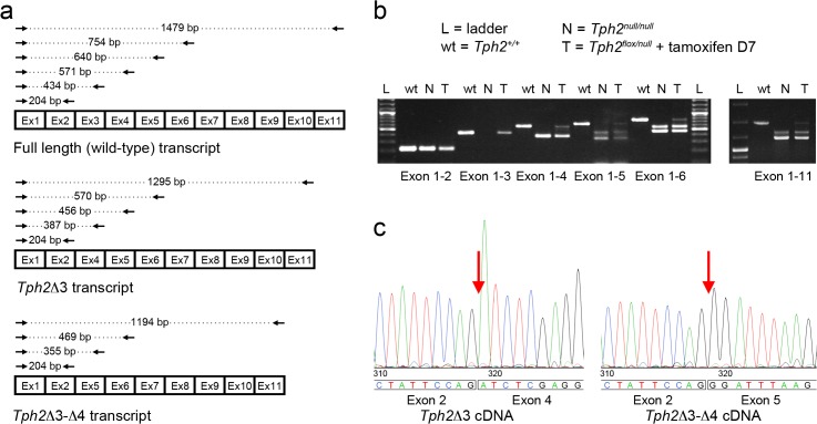
Fig 4. Characterization of Tph2 null transcripts.
(a) Schematic representation of RT-PCR products obtained from Tph2 +/+, Tph2Δ3 and Tph2Δ3Δ4 transcripts using the following primer sets: Fw1 –Rev2, Fw1 –Rev 3, Fw1 –Rev 4, Fw1 –Rev5, Fw1 –Rev6 and Fw1 –Rev11. (b) Agarose 1.5% gel showing the RT-PCR products obtained from Tph2 +/+, Tph2 null/null and tamoxifen-treated Tph2 flox/null::CMV-CreER T cDNA using the primer sets described in (a). Two distinct PCR products are obtained using Fw1 –Rev 4 (exon1-4), Fw1 –Rev 5 (exon1-5), Fw1 –Rev 6 (exon1-6), and the Fw1 –Rev 11 (exon1-11) primer sets from cDNA of Tph2 null/null and the tamoxifen treated Tph2 flox/null::CMV-CreER T mice. An additional amplicon corresponding in size to the wild-type PCR product is amplified from Tph2 flox/null::CMV-CreER T cDNA. (c) Electropherograms showing the nucleotide sequence obtained sequencing the two distinct amplicons obtained from Tph2 null/null cDNA using Fw1—Rev5 primers. Sequencing analysis shows that the second exon of the Tph2 transcript is joined to the fourth exon in the Tph2Δ3 cDNA, while it is directly connected to the fifth exon in the Tph2Δ3Δ4 transcript. L: ladder; N: cDNA from the raphe of a Tph2 null/null mice; T: cDNA from the raphe of a Tph2 flox/null::CMV-CreER T tamoxifen-treated mouse sacrificed seven days after the end of the treatment.

