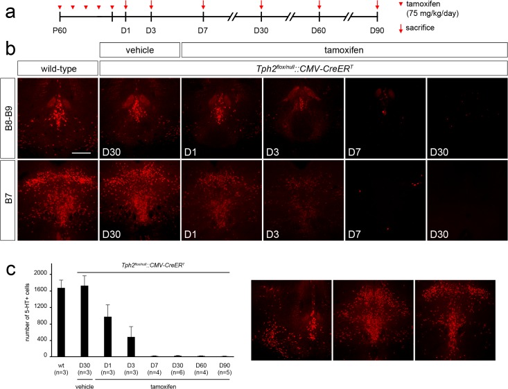
Fig 5. Tamoxifen-induced somatic recombination results in a rapid depletion of brain serotonin in adult mice.
(a) Experimental design: Tph2 flox/null::CMV-CreER T mice received tamoxifen injection once per day starting from P60 for 5 consecutive days and sacrificed 1 (D1), 3 (D3), 7 (D7), 30 (D30), 60 (D60) or 90 (D90) days after the end of the treatment, respectively. (b) Representative coronal section of B8-B9 and B7 raphe nuclei showing serotonin immunoreactivity in (from right to left) adult wild-type mice, vehicle-treated Tph2 flox/null::CMV-CreER T control mice sacrificed 30 days after the last injection, tamoxifen-treated Tph2 flox/null::CMV-CreER T mice sacrificed 1, 3, 7 and 30 days after the end of treatment, respectively. (c) Histogram showing the number of serotonin immunoreactive cells in tamoxifen-treated Tph2 flox/null::CMV-CreER T mice, as compared to wild-type and vehicle-treated Tph2 flox/null::CMV-CreER T control mice. On the right, three representative coronal sections of B8-B9 and B7 raphe nuclei showing the distinct levels where serotonin positive neurons were counted. While the number of serotonin immunoreactive cells is unchanged between vehicle-treated Tph2 flox/null::CMV-CreER T and wild-type animals, a progressive and rapid reduction of serotonin immunoreactive neurons is observed after tamoxifen treatment, resulting in brain serotonin depletion starting from 7 days after the end of the treatment. Data are presented as mean ± SEM. Scale bar: 400 μm.

