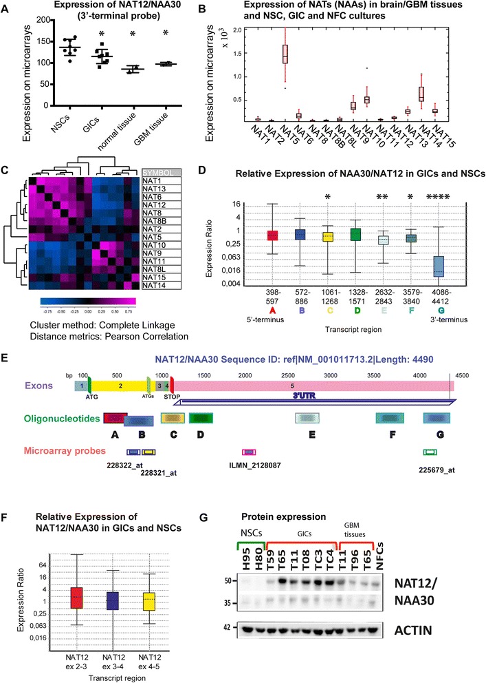
Fig. 1.

Expression of NAT12/NAA30 in GIC and NSC cultures, NFCs, and in GBM and normal brain tissues. a, Expression of NAT12/NAA30 is analysed by microarray (3’UTR Reporter = ILMN_2128087). Expression is 1.2 fold higher in NSCs than in GICs (p = 0.0281). P values were calculated using the Mann Whitney test. Due to the small sample size the values were not calculated for tissues and NFCs. b, Box and whiskers plot showing the expression of the selected NATs in all samples as measured by microarrays. c, Hierarchical clustering (with distance matrix) of the expression values for selected NAT genes in all samples. d, Expression of NAT12/NAA30 in GIC cultures was calculated using qPCR and the seven sets of oligonucleotides whose positions are delineated in (e). NSCs were used as a control. P values in this experiment were as following p(A) = 0.641, p(B) = 0.532, p(C) = 0.033, p(D) = 0.268, p(E) = 0.007, p(F) = 0.012, and p(G) = 0. The bottom and top of each box indicate the 25th and 75th percentile (the lower and upper quartiles, respectively), and the band near the middle of the box is the 50th percentile (the median). The ends of the whiskers represent the minimum and maximum of all the data. Additional details of the statistical analysis can be found in Additional file 4. e. Detailed architecture of the reference sequence of the NAT12/NAA30 transcript (NM_001011713.2) and details on probes and oligonucleotide positions. f, Relative expression of the coding regions of NAT12/NAA30 in GICs compared to NSCs as measured with Taqman probes. P values for this analysis were (0.266, 0.970 and 0.672). Additional details of the statistical analysis can be found in Additional file 4. g. Expression of the NAT12/NAA30 protein in NSC and GIC cultures, GBM and NFCs
