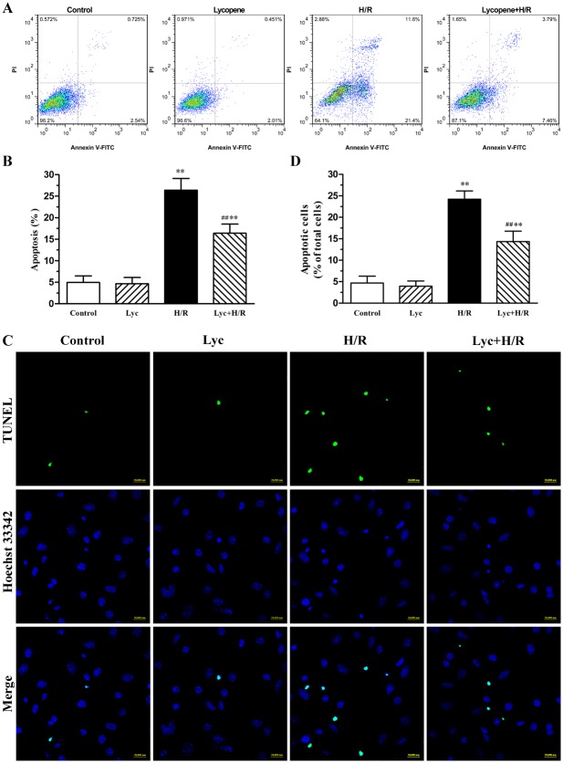
Fig 2. Effects of lycopene on H/R-induced apoptosis in cardiomyocytes.
Cardiomyocytes were pretreated with or without 5μM lycopene for 4h prior to H/R treatment. (A) Apoptotic cells were detected with flow cytometry by AnnexinV-FITC and PI counterstaining, the representative flow cytometry figures are from each group. (B) The statistical graph of apoptosis rate with AnnexinV-FITC and PI counterstaining are from three independent experiments. (C) Morphology characteristic of cardiomyocytes apoptosis was determined by staining with TUNEL and Hoechst 33342, representative images of TUNEL-positive cells (green, top row) and Hoechst counterstaining cells (blue, middle row) are shown in these representative fields. Scale bar: 20μm. (D) Quantitative analysis of TUNEL staining was from three independent experiments. The histogram shows the relative proportion of TUNEL-positive cells in the different groups. Values are mean±SEM. *P<0.05, **P<0.01 versus control; ## P <0.01 versus H/R group. (Lyc, Lycopene. H/R, hypoxia/reoxygenation).

