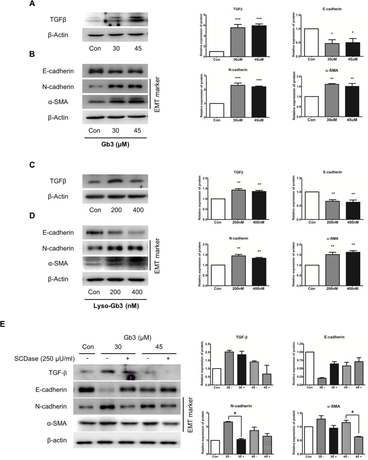
Fig 8. Effects of the expression of EMT markers by Gb3 and SCDase in SV40 MES 13 cells.
(A, B) Immunoblotting analysis of TGF-β and EMT markers following Gb3 (30 μM and 45 μM) treatment of SV40 MES 13 cells for 72 h. (C, D) Immunoblotting analysis of TGF-β and EMT markers following lyso-Gb3 (200 nM and 400 nM) treatment of SV40 MES 13 cells for 72 h. (E) The activity of TGF-β and EMT markers was assayed by the immunoblotting method treating of SCDase (250 μU/ml) and Gb3 (30 μM and 45 μM, respectively) for 72 h in SV40 MES 13 cells. Panels (A–E) show a representative experiment (right panel) and data as mean ± SEM, n = 3. (A–D) Gb3 or lyso-Gb3 vs control, (E) Gb3 vs Gb3 + SCDase, One-way ANOVA. * P < 0.05, ** P < 0.001, and *** P < 0.0001.

