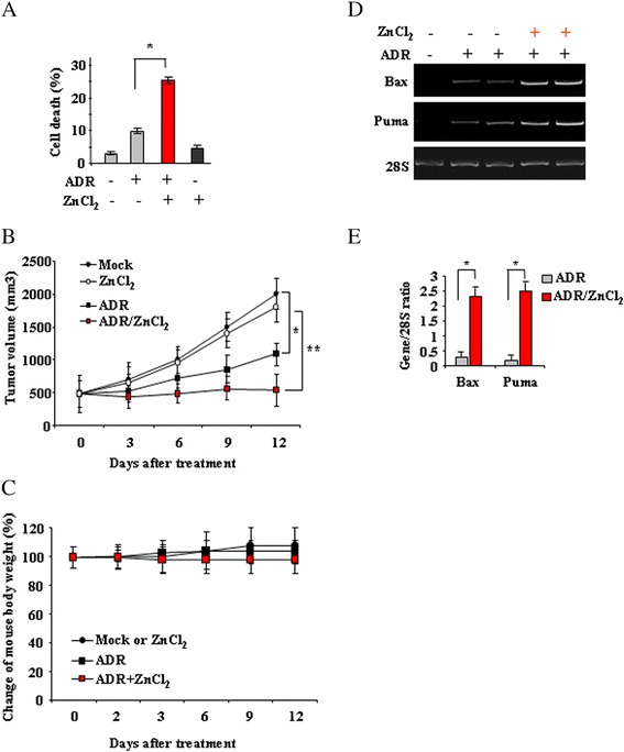
Fig. 4.

Zinc supplementation improves the antitumor effects of low-dose ADR. a RKO cells (2x10⁵) were plated at subconfluence in 60-mm Petri dish and the day after treated with ADR (0.1 μg/ml) and ZnCl2 (100 μM) alone or in combination for 24 h. Cell death was measured by trypan blue exclusion assay and expressed as percentage ± SD of three independent experiments performed in duplicate. Paired student’s t test was used for the statistical analysis of cell death percentages in the presence of ADR alone or in combination with ZnCl2 (*: P < 0.01). b RKO cells were injected i.m. into the right flanks of nude mice. Drug and ZnCl2 treatments were started when the tumors became palpable, and the size of tumors was measured twice a week and plotted as the average tumor volumes versus number of days post-treatment. Paired student’s t test was used for the statistical analysis of tumor volumes in the presence of ADR alone (black square) or in combination with ZnCl2 (red square) (*: P < 0.01). c Percentage change of mouse body weight measured throughout the experiment. d Tumors were explanted at day 12 after treatments and used for total mRNA extraction and RT-PCR analysis of p53 target genes. 28S was used as a control for efficiency of RNA extraction and transcription. RNA samples from explanted tumors show respectively one Mock tumor, two ADR-treated tumors and two ADR/ZnCl2-treated tumors. One representative experiment, out of two independent experiments generating similar results, is shown here. e Densitometric analysis of gene expression was plotted as expression ratio to 28S. The data represent the mean of two independent experiments ± S.D. Paired student’s t test was used for the statistical analysis of the normalized gene expression (Gene/28S ratios) at different ADR doses, with or without ZnCl2 (*: P < 0.01)
