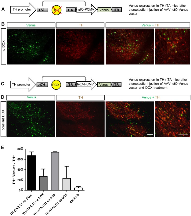
Fig 2. Cre independent adult gene expression in DA neurons of TH-tTA and TH-rtTA mice using a AAV-tetO-Venus vector.
(A) Scheme of fluorescent DA neuron labelling in non-DOX treated TH-tTA mice stereotactically injected with AAV-tetO-Venus vector in the ventral midbrain. (B) Confocal fluorescent pictures of Venus expression (green) in DA neurons co-stained for tyrosine hydroxylase (TH; red) in the substantia nigra pars compacta (SNpc) of sagittal TH-tTA mouse brain sections. (C) Scheme of fluorescent DA neuron labeling in DOX-treated TH-rtTA mice stereotactically injected with AAV-tetO-Venus vector in the ventral midbrain. (C) Scheme of fluorescent DA neuron labelling in DOX-treated TH-rtTA mice stereotactically injected with AAV-tetO-Venus vector in the ventral midbrain. (D) Confocal fluorescent pictures of Venus expression in DA neurons co-stained for TH in the substantia nigra pars compacta (SNpc) of sagittal TH-rtTA mouse brain sections. (E) Quantification reveals that in the ON-state 67% and 73% and in the OFF-state 27% and 23% of TH+ cells are also Venus-positive in TH-tTA and TH-rtTA mice, respectively. In non-transgenic mice (controls) only 5% of TH+ cells were also Venus-positive. n = 3–5. Scale bars: 250 μm and in high magnification picture 50 μm.

