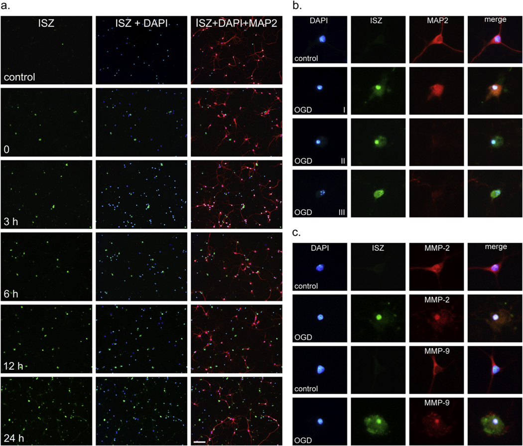
Fig. 2.
Induction of gelatinase activity and nuclear localization of MMP-2 and -9 in cortical neurons after OGD. (a) In situ zymography (ISZ) was performed on living neurons after 0, 3-, 6-, 12-, and 24-h reoxygenation as described in Experimental Procedures. ISZ-positive cells are stained green in column 1, nuclei are stained blue with DAPI in column 2, and neurons are identified with a neuron-specific marker (anti-MAP2 polyclonal antibody) in column 3. Superimposition of ISZ, DAPI, and MAP2 shows gelatinase activity in neurons. Scale bar = 100 µm. (b) The stages of gelatinase induction associated with apoptosis are shown. Stage I, early apoptosis with nuclear condensation, strong nuclear gelatinase activity, and retraction of cellular processes. Stage II, mid-stage apoptosis with further condensation of the nucleus, collapse of the cytoskeleton, and loss of MAP2 staining. Stage III, late-stage apoptosis with clear nuclear fragmentation and intense uniform gelatinase activity throughout the cell. (c) Using immunocytochemistry, MMP-2 was detected in nuclei in both control and OGD-treated cells and increased in the nucleus 24 h after OGD. MMP-9 could not be detected in the nucleus of control cells by immunocytochemistry but was detectable in neuronal nuclei after OGD.

