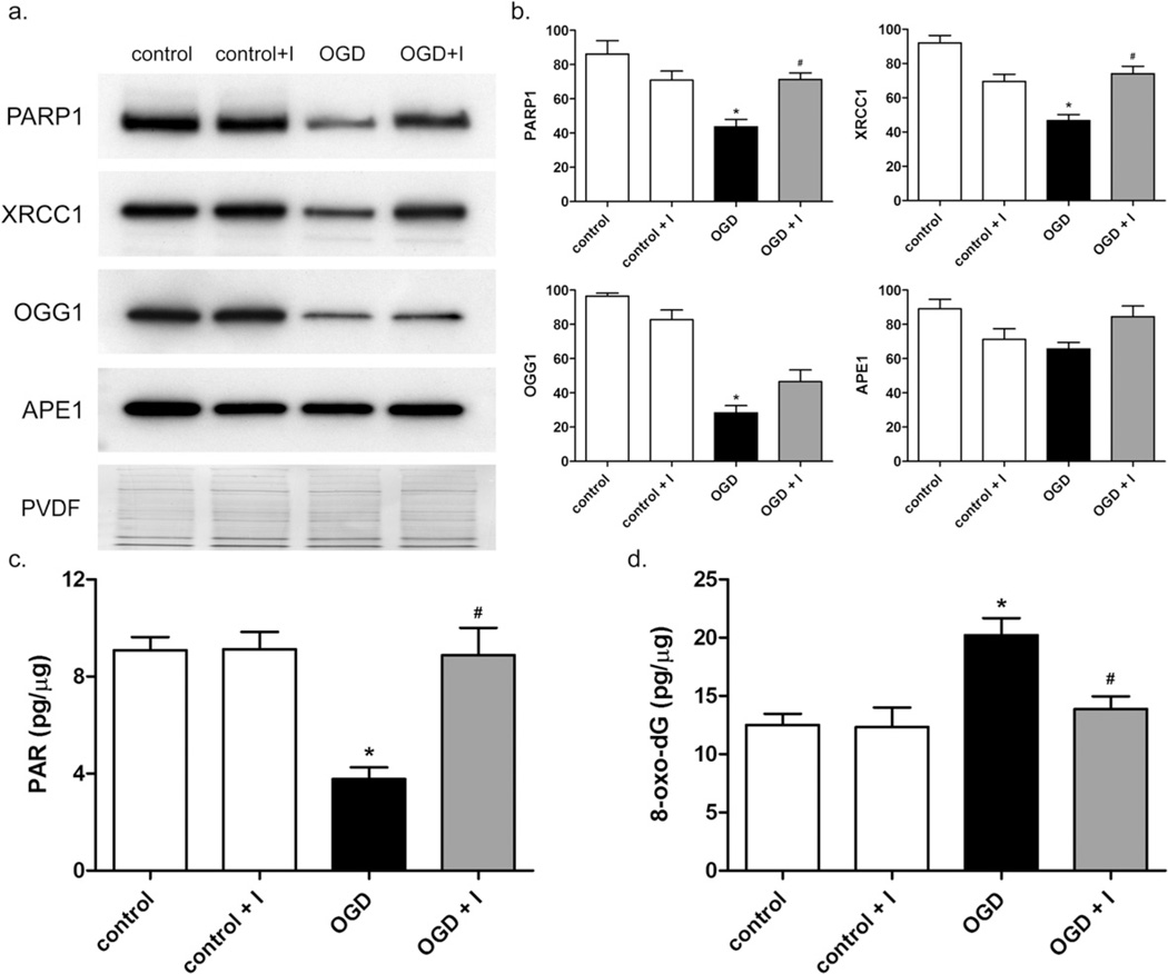
Fig. 5.
Decreased levels of DNA repair proteins and measurement of oxidative DNA damage and PARP1 activity in neuronal nuclei after OGD. (a) The levels of DNA repair proteins PARP1, XRCC1, OGG1, and APE1 in nuclear extracts of neurons exposed to control (lane 1), control plus inhibitor (lane 2), OGD (lane 3), or OGD plus inhibitor (lane 4) conditions were measured by Western blot. (b) Graphical representation of normalized relative levels of DNA repair proteins. Units are arbitrary. For PARP1 and XRCC1, protein levels were significantly lower in OGD-treated cells compared to controls (*), and significantly higher in OGD plus inhibitor compared to OGD (#), p < 0.01. OGG1 significantly decreased after OGD (*) and no significant changes in APE1 were observed. (c) Level of PAR present in the nucleus of neurons in pg PAR per µg nuclear extract. OGD caused a statistically significant decrease in nuclear PAR level compared to control cells (*), p < 0.01. In the presence of MMP-2/9 inhibitor II, a significant difference in nuclear PAR levels between OGD and OGD plus inhibitor was observed (#), p < 0.01. (d) After OGD and 24-h reoxygenation, 8-oxo-dG in genomic DNA (pg per µg DNA) was measured as described in Experimental Procedures. 8-oxo-dG was significantly elevated in OGD-treated cells compared to controls (*), p < 0.01, and significantly lower in OGD plus inhibitor compared to OGD (#), p < 0.05. Results are means and standard deviations from 4 independent experiments.

