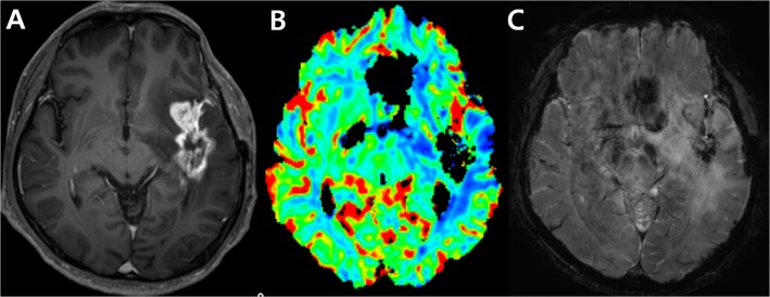
Fig 2. An example of an uninterpretable DSC perfusion MR image. Images obtained in a 56-year-old man clinicoradiologically considered as having tumor progression.
Contrast-enhanced, T1-weighted image (A) acquired 19 weeks after concomitant chemoradiotherapy (CCRT) shows an enhancing lesion in the temporal lobe. DSC perfusion MR image (B) shows signal loss in the corresponding contrast-enhancing lesion due to treatment-related hemorrhage confirmed on susceptibility weighted image (C).

