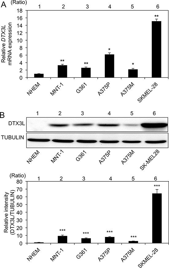
Figure 2. Increased expression levels of DTX3L in melanoma cells in humans.

Expression levels (means ± SD) of DTX3L transcript A. and protein B. in normal human epithelial melanocytes (NHEMs) and melanoma cells (MNT-1, G361, A375P, A375M and SK-Mel28) determined by real-time PCR A. and immunoblot B. analyses are presented. Expression levels of α-TUBULIN protein are presented as an internal control B. Expression levels of Dtx3l (means ± SD) determined by real-time PCR A. and densitometric analyses of the bands B. in 3 independent experiments are presented as graphs showing relative values (lanes 2–6) for NHEMs (lane 1). * and **, Significantly different (*, p < 0.05; **, p < 0.01) by Dunnett's test.
