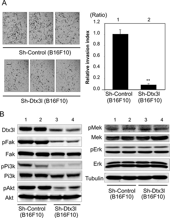
Figure 4. Decreased cell invasion of Dtx3l-depleted murine melanoma cells.

Matrigel-invasion assay was performed with control (Sh-Control) and Dtx3l-depleted (Sh-Dtx3l) B16F10 murine melanoma cells A. Photographs of cells invading the membrane stained with hematoxylin are presented (left). After invading cells had been counted in five random microscopic fields in each Matrigel-invasion assay, the results of 3 independent assays were normalized and are presented as an invasion index (right). Expression (Dtx3l, Fak, Pi3k, Akt, Mek and Erk) and phosphorylation (pFak, pPi3k, pAkt, pMek and pErk) levels in two kinds of DTX3L-depleted (Sh-DTX3L) and control (Sh-Control) murine melanoma cells determined by immunoblot analysis are presented B. Expression levels of α-Tubulin protein are presented as an internal control B. Significantly different (**, p < 0.01) from the control (Sh-Control) by Student's t-test. Scale bar, 50 μM.
