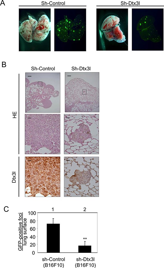
Figure 6. Decreased lung metastasis in Dtx3l-depleted melanoma cells in vivo.

Results of morphologic analysis of lung metastasis of control (Sh-Control) and Dtx3l-depleted (Sh-Dtx3l) murine B16F10 melanoma cells injected into tail veins of nude mice A. are presented. Animals were dissected to observe lung metastases at 14 days after inoculation. Lung metastases were macroscopically visualized by GFP fluorescence images. Metastatic foci derived from control (Sh-Control) and Dtx3l-depleted (Sh-Dtx3l) cells B. were microscopically confirmed by low (top panels in B) and high (middle and bottom panels in B) magnification of HE staining (HE) and immunohistochemistry (Dtx3l). Number of GFP-positive metastatic foci per lung surface C. after inoculation of control (Sh-Control; n = 4) and Dtx3l-depleted (Sh-Dtx3l; n = 4) murine B16F10 melanoma cells is presented. Significantly different (**, p < 0.01) from the control (Sh-Control) by the Student's t-test. Scale bar, 200 μM (low magnification) and 25 μM (high magnification).
