
Figure 5. Combination treatment induced PUMA-dependent apoptosis.
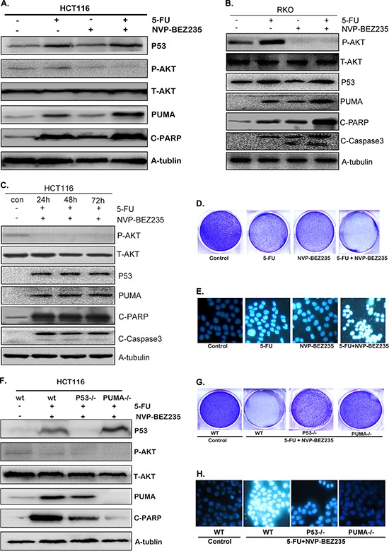
A–C. Western blotting showing the expression of p53, P-Akt(S473), PUMA, cleaved PARP and Caspase3 after the treatment of 200 uM 5-FU, 400 nM NVP-BEZ235 or their combination for (A) 12 hours in HCT-116 cells, (B) 12 hours in RKO cells, (C) 24, 48 or 72 hours in HCT-116 cells. (D and G). Colony formation of HCT-116 cells. Cells were treated with different drugs for 24 hours, followed with crystal violet staining of attached cells at 14 days. (D) 200 uM 5-FU, 400 nM NVP-BEZ235, or their combination in wild-type HCT-116 cells, (G) combination treatment in wild-type, p53−/− or PUMA−/− HCT-116 cells. E. and H. Hoechst 33342 morphological examination of apoptosis in HCT-116 cells, (E) treated with 200 uM 5-FU, 400 nM NVP-BEZ235, or their combination for 12 hours in wild-type HCT-116 cells, (H) combination treatment for 12 hours in wild-type, p53−/− or PUMA−/− HCT-116 cells. F. The expression of p53, P-Akt, PUMA and cleaved PARP were detected in wild-type, p53−/− or PUMA−/− HCT-116 cells. Similar results were obtained from three independent experiments.