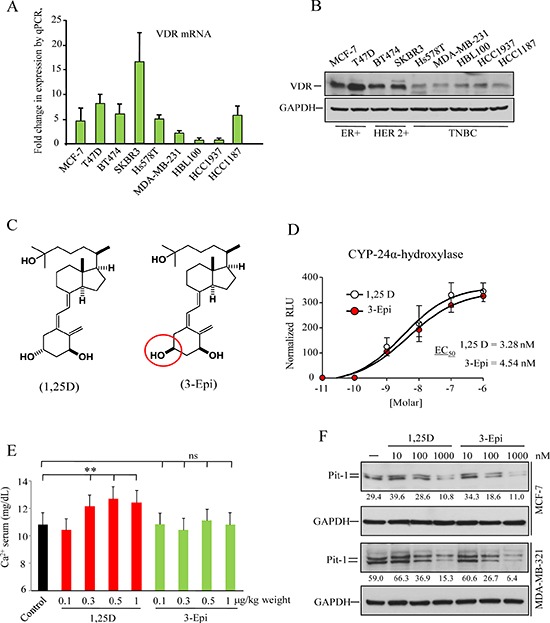
Figure 3. Vitamin D receptor (VDR) expression in human breast cell lines, and biological activity of the vitamin D derivative 1α, 25-dihydroxy-3-epi-vitamin D3 (3-Epi).

A. Real-time PCR of VDR mRNA in the human breast tumor cell lines MCF7, T47D, BT474, SKBR3, Hs578T, MDA-MB-231, HBL100, HCC1937, and HCC1187. VDR expression are plotted as mean ± SD of triplicates using 18S gene expression as control. B. Western blot of VDR protein from the breast tumor cell lines described above. C. Structure of 1, 25D and 3-Epi. D. Transcriptional activation of the 24-hydroxylase gene (CYP24A1) by 1, 25D and 3-Epi. MCF-7 cells were transfected with the pCYP24A1-luc vector (encoding the luciferase gene under control of a consensus vitamin D response element) and treated with 1, 25D or 3-Epi (10−11 to 10−6 M) for 24 h. The EC50 values derived from dose-response curves, and represent the analogue concentration capable of increasing luciferase activity by 50%. Error bars represent standard deviation (SD). E. Serum calcium level in mice treated with 1, 25D and 3-Epi. Five mice per group were treated with 0.1, 0.3, 0.5, and 1 μg/kg weight of 3-Epi, 1, 25D, or vehicle (sesame oil) every other day for 3weeks, and calcium levels were measured on day 21. Error bars represent standard deviation (SD). ** = P < 0.01, ns = not significant. F. MCF-7 and MDA-MB-231 cells were treated with 1, 25D and 3-Epi (10−8 to 10−6 M) for 48 h. Representative immunoblot of Pit-1 and GAPDH. Values correspond to a densitometric analysis.
