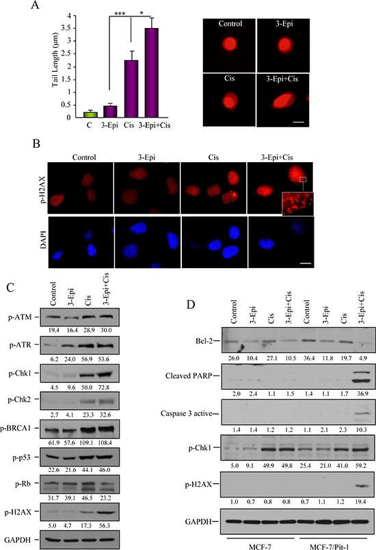
Figure 6. 3-Epi enhances cisplatin effect in Pit-1-sensitized breast cancer cells.

A. Pit-1 overexpressing MCF-7 cells (MCF-7/Pit-1) treated with ethanol, 100 nM of 3-Epi, 5 μM of cisplatin, and 100 nM of 3-Epi + 5 μM of cisplatin were analyzed for DNA-damage in a comet assay. DNA-damage induced by each treatment is indicated by average tail length. Data are represented as mean ± SD. A representative image is shown in B. Scale bar: 10 μm. B. Immunofluorescence of p-H2AX protein, as marker of DNA-damage, after treatment with ethanol, 3-Epi, cisplatin, and 3-Epi+cisplatin in MCF-7/Pit-1 cells. Scale bar: 10 μm. Cisplatin and 3-Epi+cisplatin modify DNA-damage phosphorylated proteins. C. Immunoblot analysis of extracts in the conditions described above was done for the DNA-damage phosphorylated (p) proteins, p-ATMSer1981, p-ATRSer428, p-Chk1Ser296, p-Chk2Thr68, p-BRCA1Ser988, p-p53Ser15, p-RbSer139, and p-H2AXSer139. GAPDH was used as loading control. Values correspond to a densitometric analysis. D. Immunoblot analysis of Bcl-2, cleaved PARP, caspase 3 active, p-Chk1Ser296, p-H2AXSer139, and GAPDH (used as loading control) in MCF-7 and MCF-7/Pit-1 cells treated with ethanol, 100 nM of 3-Epi, 5 μM of cisplatin, and 100 nM of 3-Epi + 5 μM of cisplatin. Values correspond to a densitometric analysis.
