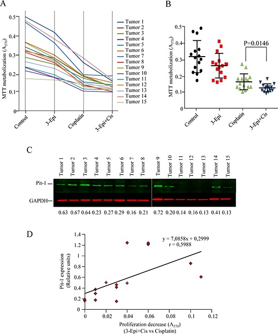
Figure 8. Cell proliferation response to 3-Epi+cisplatin treatment in primary cultures of breast tumors is related to Pit-1 levels.

A. Fifteen primary cultures of human breast tumors (BT) were treated for 48 h with ethanol (controls), 100 nM of 3-Epi, 5 μM of cisplatin, or 100 nM of 3-Epi + 5 μM of cisplatin. Then, MTT was added and absorbance measured at 570 nm. Absorbance values are plotted as the mean of quadruplicate values. B. Cell proliferation response in primary breast cultures was significantly (P = 0.014) reduced after 3-Epi+cis as compared with cisplatin. C. Representative Western blot of Pit-1 levels in primary breast tumors. Pit-1 expression was evaluated by quantitative Western blot. Values were corrected by GAPDH expression. D. Statistical analyses indicated that reduced proliferation response to 3-Epi+cis as compared to cisplatin alone is correlated with Pit-1 levels.
