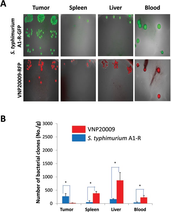
Figure 3. Comparison of tissue distribution of S. typhimurium A1-R and VNP20009 in tumor and normal tissues.

When the average tumor volume reached approximately 70 mm3, S. typhimurium A1-R (1×10⁷ CFU) or VNP-20009 (1×10⁷ CFU) were injected into the tail vein. Tissues were removed 6 days after bacteria administration. Bacteria were isolated from the tumor and organs and cultured in LB agar (*p<0.05)
