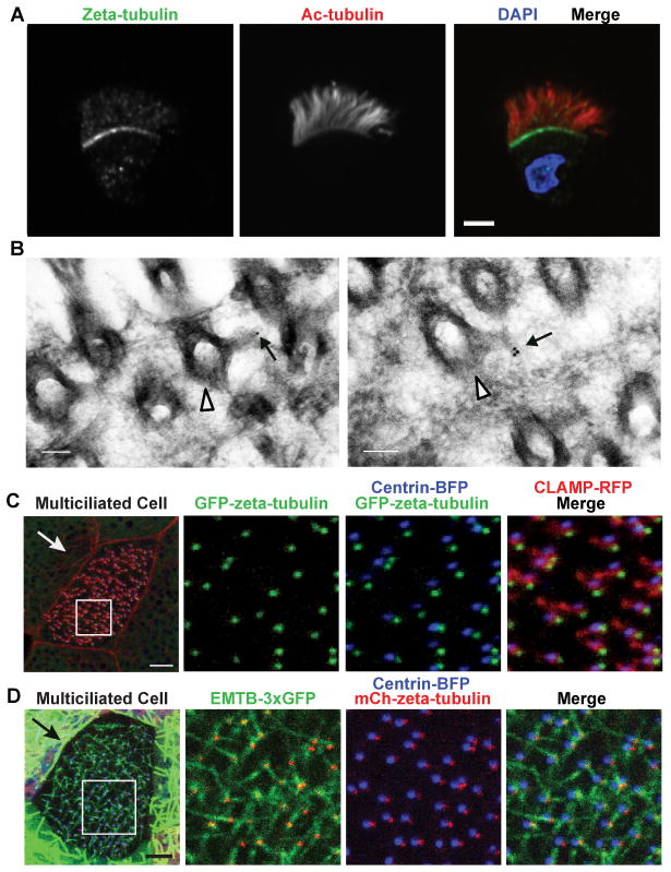
Figure 2. Zeta-tubulin localizes to the basal foot in multiciliated cells.
A) Dissociated Xenopus multiciliated oviduct cells stained for zeta-tubulin (green), acetylated tubulin (red), and DAPI (blue). Scale bar, 5 μm. B) Transmission EM of oviduct tissue stained with zeta-tubulin antibody and 10 nm gold-conjugated secondary antibody. Arrowhead indicates basal body, and arrow indicates labeling of basal foot. Scale bars, 100 nm. C) Confocal image of live tadpole epidermal multiciliated cell expressing GFP-zeta-tubulin (green), CLAMP-RFP (red), and centrin-BFP (blue). Arrow shows direction of ciliary beating. Scale bar, 5 μm. D) Confocal image of live tadpole epidermal multiciliated cell expressing EMTB-3XGFP (green), mCherry-zeta-tubulin (red), and centrin-BFP (blue). Arrow shows direction of ciliary beating. Scale bar, 5 μm. See also Figures S2 and S3.

