
Figure 1.
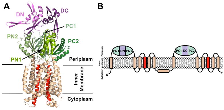
(A) Cartoon representation of monomeric CusA (PDB code: 4DNT) showing the docking, porter, and transmembrane subdomains. The N- and C-terminal docking (DN and DC) and porter (PN1, PN2, PC1, and PC2) subdomains are colored shades of purple and green, respectively, while the transmembrane subdomain is colored wheat, except for the central transmembrane helices, TM4 and TM10, which are colored red. (B) RND transporter membrane topology with two periplasmic domains, each containing two porter subdomains and one docking subdomain. An additional extracytosplasmic α-helix between TM6 and TM7 is located near the cytoplasmic membrane surface and runs almost parallel to it. Subdomain color designation is as in (A).