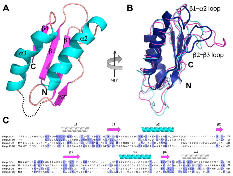
Figure 2.
(A) Cartoon representation of MmpL11-D2 structure with missing residues 479 – 489 depicted as dashed lines. α-helices, β-strands, and loops are colored cyan, magenta, and wheat, respectively (B) MmpL11-D2 colored blue, is rotated 90° clockwise from (A) and structurally aligned with RND PC1 porter subdomains from ZneA (PDB code: 4K0E) and MexB (PDB code: 2V50), colored magenta and cyan, respectively. (C) MmpL11 and MmpL3 Cluster II D1 and D2 domain sequence alignment based on secondary structural prediction and MmpL11-D2 structure. Cylinders (α-helices) and arrows (β-sheets) colors correspond to the secondary structural elements in (A). The predicted α-helices (α1 and α4) are shown as white cylinders.

