
Figure 3.
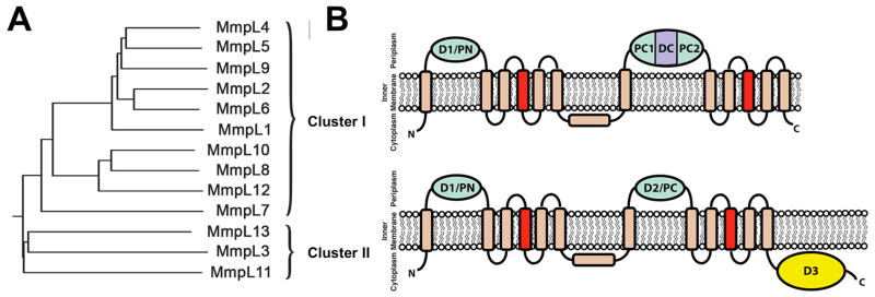
(A) Phylogenetic tree of MmpL proteins reveals two distinct clusters where the (B) predicted membrane topologies of MmpL Clusters I and II proteins are based on RND transporters. The predicted porter domains (N-terminal D1 (PN) and Cluster I C-terminal D2 (PC1 and PC2) and Cluster II C-terminal D2 (PC)) are colored green and the predicted Cluster I C-terminal docking domain (DC) is colored purple. D3 is colored yellow while the transmembrane subdomain is colored wheat, except for the central transmembrane helices, TM4 and TM10, which are colored red. The predicted additional extra-cytoplasmic α-helix located between TM6 and TM7 is shown almost parallel to the cytoplasmic membrane surface, as observed in RND transporter structures (Fig. 1B).