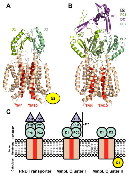
Figure 6.
Phyre2 models of (A) MmpL11 with additional restraints from the crosslinking results and (B) MmpL4. The transmembrane domains are colored wheat, except for TM4 and TM10, which are colored red. The different periplasmic porter subdomains are in shades of green and the proposed MmpL4 docking domain is colored purple. The Cluster II MmpL (MmpL11) cytoplasmic D3 domain is signified by a yellow circle. (C) A cartoon representing the domain architecture of RND transporters, MmpL Cluster I and II proteins. Subdomain color designations are as in (A) and (B).

