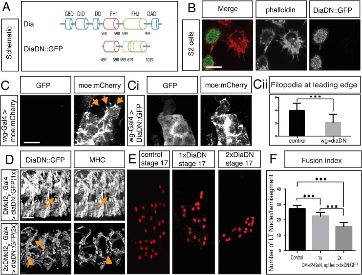
Fig 3. Diaphanous is required for myoblast fusion.
A. Schematic diagram of Dia domain structure and a deletion construct that renders Dia dominant negative (DiaDN). DiaDN consists of the FH1 domain and a partially deleted FH2 domain; the deleted aa 750–770 in the FH2 domain is indicated by the dashed area. B. Expression of DiaDN reduces filopodia number in S2R+ cells. S2R+ cells that were transfected with DiaDN::GFP (green in merge, grey in single channel) have less filopodia-like, protrusive structures (phalloidin, red in merge, grey in single channel) relative to untransfected cells (n = 20, p<0.001). Scale bar: 10μM. This reduction was rescued by expression of Dia::GFP (S3C Fig). C-Ci. UAS-diaDN::GFP was expressed in leading edge cells using wg-Gal4. Moesin::mCherry was also expressed in leading edge cells to visualize actin. In stage 15, GFP-negative control cells, filopodia structures are seen (C, arrow). DiaDN::GFP significantly reduced filopodia formation (Ci). Scale bar: 2.5μM. Cii. Filopodium number was quantified in each wg-Gal4 expressing stripe. DiaDN significantly reduced filopodia formation in leading edge cells relative to control (2.1±1.29μM vs 3.95±1.15μM, p<0.001). D. Increasing DiaDN concentration in myoblasts through higher temperature and genetic copy number leads to an increased fusion block. Three hemisegments of a lateral view of a stage 16 embryos stained with GFP and MHC antibody are shown. Myoblast fusion is relatively normal in 1xDMef2-Gal4>1xdiaDN::GFP embryos at 29°C (upper panel), with few free myoblasts (arrow). In 2xDMef2-Gal4>2xdiaDN::GFP embryos (lower panel), a higher degree of fusion block (arrows) and muscle detachment (arrowheads) are observed. E. One hemisegment of stage 17 embryo showing apME-NLS::dsRed labeled nuclei in the four lateral transverse (LT) muscles: From left to right: apME-NLS::dsRed labeled nuclei in stage 17 LT muscles in control, in 1xDMef2-Gal4> 1xUAS-diaDN::GFP, and in 2xDMef2-Gal4> 2xUAS-diaDN::GFP embryos F. Fusion index of Stage 17 lateral transverse (LT) muscles confirms the degree of fusion block in DiaDN embryos. In control embryos, 27.1±2.3 nuclei were counted in each hemisegment (n = 40 hemisegments). 1xDMef2-Gal4> 1xUAS-diaDN::GFP reduces the number of dsRed positive nuclei in each hemisegment to 22.5±2.2 (p<0.001), whereas 2xDMef2-Gal4> 2xUAS-diaDN::GFP further reduces the number to 15.5±2.7(p<0.001). Scale bar: 24μM.

