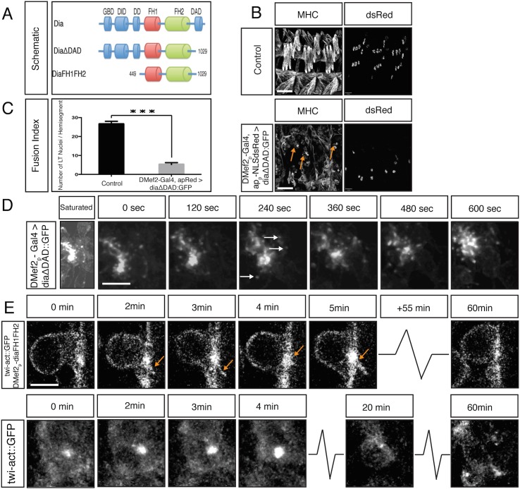
Fig 5. Constitutively active Dia blocks myoblast fusion.
A. Schematic diagram of Dia domain structure and different constitutively active Dia deletion constructs used in this study. B. Whole mount lateral view of three hemisegments from stage 16 embryos showing the (MHC) labeled muscles and nuclei (apME-NLS::dsRed) of the lateral transverse (LT) muscles. Scale bar: 24μM. Expression of DiaCA blocks myoblast fusion as visualized by many free myoblasts (arrows). This fusion defect is not due to a failure in FC specification as witnessed by expression of apME-NLS::dsRed in nuclei. C. Fusion index confirms a total block in myoblast fusion: dsRed positive nuclei in LT muscles/ hemisegment were counted in control (26.6±1.5) and DMef2-Gal4>UAS-diaΔDAD::GFP (5.2±1.0) (n = 40 hemisegments/genotype) (p<0.001). D. Dynamics of DiaΔDAD::GFP expression in myoblasts. Still images from time-lapse of a stage 14 DMef2-Gal4>UAS-diaΔDAD::GFP embryo. Saturated image shows outline of cells and is used to localize myoblasts attempting to fuse. Filopodia-like protrusions undergo highly dynamic extension and retraction at areas of cell contact. DiaΔDAD::GFP localizes at the tip of those protrusions (arrows), and this signal moves as the filopodium extends and retracts (S3 Movie). Scale bar: 5μM E. Still images from time-lapse of stage 14 twist-actin::GFP; DMef2-Gal4>UAS-diaFH1FH2 embryo. Images at 0 and 3 min show filopodia-like protrusions (arrows) emanating from the FCM, which adheres to the FC but is unable to fuse. The Actin::GFP signal is enriched at the fusion site during the entire time lapse sequence (1 hr). Compare to control FCM (twist-actin::GFP, lower panel), which fuses with FC in 30 minutes. Scale bar: 4μM

