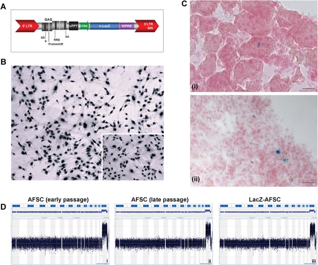
Fig 2. Labelling, in vivo tracking and genomic stability testing of amniotic fluid derived renal stem cells.

(A) Schematic representation of the lentiviral transfer construct. The nuclear localized LacZ reporter gene (n-LacZ) is driven by the chicken beta-actin promoter (CBA) and is followed by the woodchuck hepatitis post-transcriptional regulatory element (WPRE). The promoter is preceded by the central polypurine tract (cPPT) and the LV cassette is flanked by the 5′ long terminal repeat (LTR) and 3′ self-inactivating (SIN) LTR. (B) In vitro β-galactosidase staining after stable transduction. (C) In vivo engraftment of n-LacZ-cells with a representative figure of the kidney after 6h and the lung after 24h. This image does not suggest engraftment of the cells in the target organ, merely their presence. (D) CGH dataset displays the 60K genome-wide array results for a hAFSCs cell line with renal phenotype at passage 14 (left), at passage 30 (middle) and after lentiviral manipulation (right).
