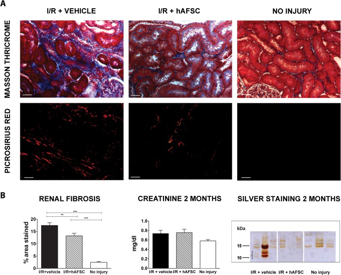
Fig 5. Long-term preservation of renal function and inhibition of tubulo-interstitial fibrosis induced by stem cell therapy.
(A) Histochemical staining of representative kidney sections from different experimental groups using Masson’s and PicroSirius at 2 months after injury for the three cohorts. (B) Quantitative analysis of the presence of renal fibrosis depicted on the left for the three cohorts (vehicle treated (black), hAFSC treated (striped) and no injury (white)). The middle panel shows the serum creatinine values for the three groups (no statistically significant difference). The right panel shows a representative image of a silver staining procedure of urine at 2 months (n = 3 per group). The group with renal reperfusion injury shows the presence of micro-proteinuria, which was absent in both other groups. * p<0.05, ** p<0.01 *** p<0.001. (scale bar = 20 μm)

