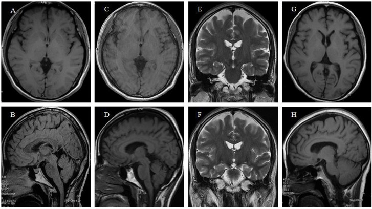
Fig 2. Brain MRI and CT scans of two patients of the family.
A,B: brain MRI scan of the proband at the age of 42; C-F: brain MRI scan of the proband at the age of 44; G,H: brain MRI scan of the patient Ⅳ20 at the age of 46. Atrophy of whole brain was present in both patients. The frontotemporal atrophy was the most noticeable, while in the proband the hippocampus was relatively well-preserved.

