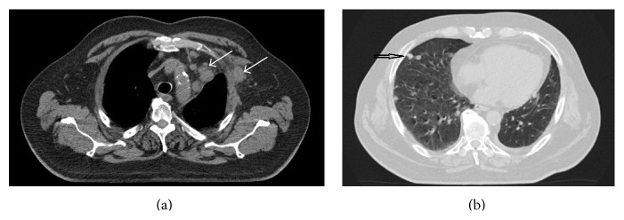
Figure 2.

(a) Computed tomography scan of the thorax showing left-sided axillary nodal metastasis and mediastinal lymph node metastases. Status before palliative radiotherapy. (b) Computed tomography scan of the thorax showing right-sided pulmonary metastases (example of the patient's bilateral metastases). During radiotherapy, this region received a cumulative dose of <1 Gy.
