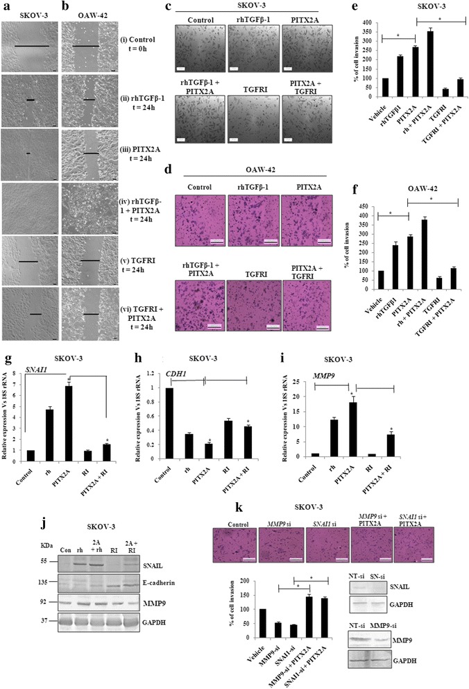
Fig. 4.

The regulation of invasion and migration is served by PITX2-mediated activation of TGF-β signalling pathway. Wound healing assay was performed of SKOV-3 (a) and OAW-42 (b) cells after treatment with rhTGFβ1 (ii), transient transfection of PITX2A (iii) and treatment with rhTGFβ1 (iv) or TGFRI (v) of PITX2-transfected cells in the time course of 24 h. T = 0 h at control cells (i) signifies the time of scratching the cells with pipette tips. The arrows indicate the width of wound and the assay was repeated three times independently. Scale bar: 50 μm. Transwell migration and invasion assay was performed in SKOV-3 (c) and OAW-42 (d) cells after treatment and/or transient transfection as mentioned. Scale bar: 200 μm. Cells at three independent fields for each well were counted and plotted with error bar for SKOV-3 (e) and OAW-42 (f) cells. g-i pcDNA3 or PITX2A-transfected SKOV-3 cells were treated with rhTGFβ1 (10 ng/ml) or TGFRI (20 ng/ml) followed by isolation of RNA and Q-PCR with the primers of SNAI1 (g), CDH1 (h) and MMP9 (i). Relative gene expression is indicated as ‘fold’ change in the Y-axis (mean ± SEM). * represents p < 0.01. j Lysates of the cells transiently transfected and/or treated as indicated were immunoblotted with respective antibodies and the representative gel image was shown. k Transwell invasion assay was performed with SKOV-3 cells after transient transfection as mentioned (top). Cells at three independent fields for each well were counted and were plotted with error bar (bottom). The efficiency in knocking down the expression of SNAIL and MMP9 proteins by SNAI-(SN)-si and MMP9-si respectively was verified by Western blot of the transfected cell lysates with respective antibodies. Here, GAPDH was used as loading control (bottom)
