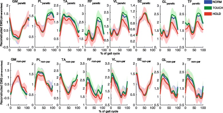
Fig. 4.

Reconstructed time normalized EMG patterns (dimensionless) of paretic (upper row) and non-paretic (lower row) leg, based on the three modes for each condition, averaged over strides and participants. Using the time courses Y (j)k we reconstructed signals as superposition . We further added the DC-values of the original EMGs to these time courses to generate EMG-like patterns. Solid lines indicate averages, shaded areas indicate SD over participants. Stride cycle starts and ends with initial contact of the nonparetic leg. GM = m. Gastrocnemius medialis; TA = m. Tibialis anterior; PL = m. Peroneus longus; RF = m. Rectus femoris; VL = m. Vastus lateralis; ST = m. Semitendinosus GL = m. Gluteus medius; TF = m. Tensor fascia latae
