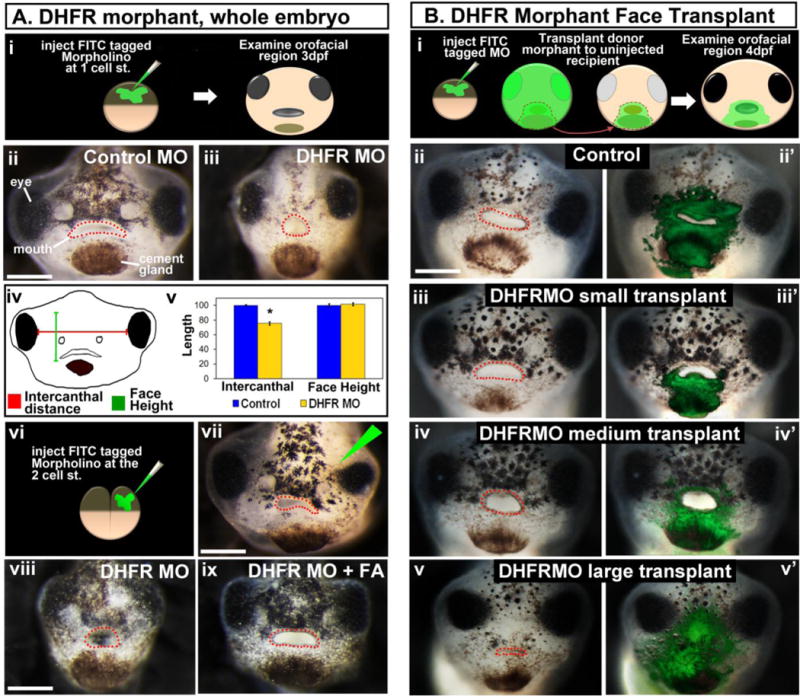
Figure 2.

DHFR morpholino knockdown results in abnormal orofacial development. A) Whole DHFR morphant analysis. i) Schematic showing the experimental design. ii,iii) Representative frontal views of faces injected with control (ii) or DHFR (iii) morpholinos. iv) Schematic of facial dimensions measured. v) Bar graph showing quantification of facial dimensions normalized to the controls (set to 100), asterisk designates statistical significance for 2 experiments (n=60) p value < 0.001. vi) Schematic showing the experimental design for vii. vii) Representative frontal view of the face of an embryo injected in one cell as shown in vi. The injected side is indicated by the green triangle. viii, ix) rescue experiment showing representative frontal views of faces injected with DHFR MO (viii) or DHFR MO and treated with folinic acid (FA) (ix). B) Localized DHFR morpholinos using face transplants. i) Schematic of the experimental design. ii–v’) Representative frontal views of embryos that received DHFR morphant tissue. Prime roman numerals are composites of the unprimed counterpart with fluorescent overlays showing the location of morpholino containing tissue (n= 2 experiments, n=11). All embryos were imaged at same magnification, scale bar = 200 μm. The mouth is outlined in red dots.
