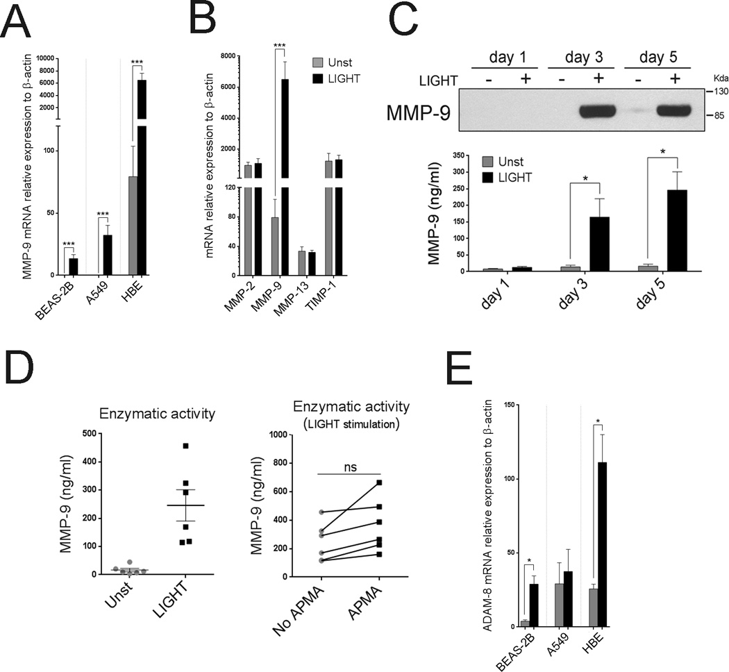
Figure 4. LIGHT promotes selective expression of MMP-9 and ADAM-8 in lung epithelial cells.

(A) Mean MMP-9 mRNA levels after 5 days in ECs cultured with (black bars) or without (gray bars) rLIGHT. n=3 independent experiments. (B) Mean MMP-2, MMP-9, MMP-13, and TIMP-1 mRNA relative expression, in HBE cells after 5 days of culture. n=6 independent experiments. (C) MMP-9 protein in supernatants of cultured HBE cells over time, by immunoblotting (top). Enzymatic activity of secreted MMP-9 over time measured by enzymatic fluorometric assay (bottom). Data representative of three independent experiments. (D) Enzymatic activity of secreted MMP-9 in HBE culture supernatants after 5 days, from 6 individual donors, measured by enzymatic fluorometric assay. Endogenous versus potential activity shown on the right after APMA activation. (E). Mean ADAM-8 mRNA levels after 5 days of culture in the absence (gray bars) or presence of LIGHT (black bars). n=3 independent experiments.
