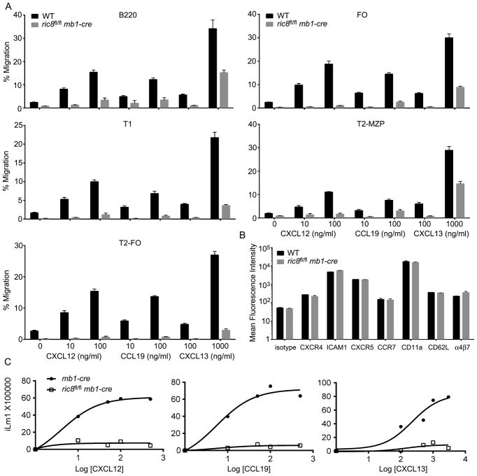
Figure 3.
B cell specific loss of Ric-8A impairs responses to chemokines. (A) Chemotaxis assays using splenic B cells from mixed bone marrow chimera mice. C57BL/6 CD45.1 mice were reconstituted with bone marrow from ric8fl/flmb1-cre CD45.2 mice and C57BL/6 CD45.1 mice. Eight weeks later purified B220+ cells were immunostained for B cell subset markers, CD45.1, and CD45.2; and subjected to chemotaxis assays with the indicated concentrations of chemokine. The percentages of migratory cells for each subset and each genotype are shown. The data is from 4 reconstituted mice performed in duplicate. The indicated chemokine concentrations are ng/ml. Data shown as mean +/− SEM. (B) Flow cytometry analysis of CXCR4, ICAM1, CXCR5, CCR7, CD11a, CD62L and α4β7 expression on B220+ cells from the same mice used in part A. WT and ric8fl/flmb1-cre cells were distinguished by CD45.1 versus CD45.2 immunostaining. (C) Intracellular calcium response to chemokines using B cells from mb1-cre and ric8fl/flmb1-cre mice. The cells were stimulated with indicated amount of CXCL12, CCL19 or CXCL13 and the induced change in intracellular calcium monitored over 3 minutes. Data represents the maximal calcium change plotted against the chemokine concentration (ng/ml). Data are from 3 experiments.

